Recording a 12 lead ECG/EKG
A 12 lead ECG/EKG (electrocardiogram) is used to record the electrical activity of the heart. Some advanced first responders may be trained in recording 12 lead ECG/EKGs.
Electrical activity of the heart
Electrical impulses start at the sino-atrial node in the heart. The impulse travels through adjacent fibres, to the atrio-ventricular node, then along the bundle of His which sits in between the two ventricles, before it spits into the bundle branch fibres (right and left).
The left bundle branch splits again into the left anterior hemi-fascicle and the posterior hemi-fascicle. The bundles terminate eventually in the Purkinje fibres. As the impulse passes, the cardiac muscle fibres contract, first atria, then ventricles, and a full heartbeat occurs.
Characteristics of the ECG/EKG 
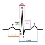
– P wave is the ECG display of atrial depolarisation and contraction
– QRS complex is the ventricular contraction
– T wave is repolarisation of the ventricles i.e. their return to a resting state
– Q wave is the first part of the QRS complex, but only if it goes downward
– R wave is the upward deflection of the QRS complex whether there is a q wave or not
– S wave follows R wave and is called as such as long as the S wave dips below the baseline.
ECG timing
– 5 large squares per second, 300 large squares per minute, so one large square is 0.2 seconds.
– 1 small square is 0.04 seconds.
– Heart rate of 60 is one QRS complex every 5 squares.
– The PR interval, measured from start of p wave to beginning of QRS, should be 0.12 – 0.2 seconds ie 3-5 small squares.
– QRS is normally 0.12, or 3 small squares.
ECG procedure
The patient will ideally be still, as skeletal muscle movement will be picked up on the ECG. The lead must be applied in the correct places, with adherence to the skin maximised by shaving the areas, enough for the lead to be applied.
There will usually be 10 leads.
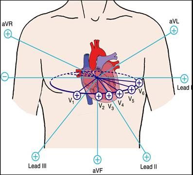
Leads are named. They are V1, V2, V3, V4, V5, V6 to be applied over the thorax, and four limb leads to be applied usually at each wrist and each media lower leg. These are usually labelled LA (left arm), RA (right arm), LL (left leg), RL (right leg).
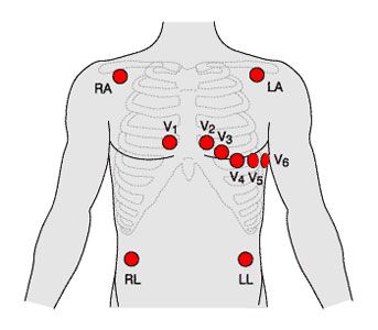
The 12 parts of the ECG are labelled V1, V2, V3, V4, V5, V6, VR, VL, VF, I, II,III.
The V leads record activity from the corresponding region of the heart that they sit next to. For example V1 sits to the right of the sternum at about 4th intercostals space. That is over the right ventricle. V2 is on the opposite side of the sternum. V3 is lower, and lateral to V2, corresponding to the septal region of the ventricles, and so on, up to the left ventricle recorded by V6.
The last 6 of those leads look in a vertical plane at the heart. VR records the electrical activity from the right side ie the right atrium, VL from the left, VF from the base. I and II record information from the vertical plane in between left vertical and the base of the heart, and lead III in between right vertical and the base of the heart